
Introduction
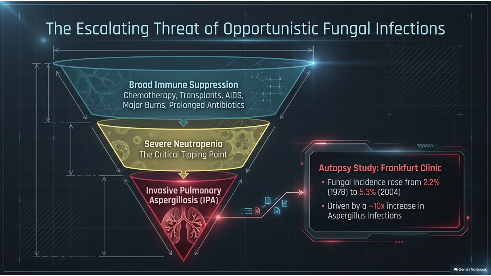
Invasive pulmonary aspergillosis is a severe form of invasive fungal infection that can develop in patients with significantly reduced immunity. The condition is particularly important in patients receiving cytostatic or immunosuppressive medications, patients undergoing transplantation, and individuals with other conditions associated with severe immune suppression.
The development of modern medical technologies has improved the diagnosis and treatment of severe diseases, including oncological and oncohematological disorders. However, treatments that suppress the immune system can increase the risk of opportunistic infections. Patients with AIDS, complications following major abdominal surgery, severe burns, premature newborns, and patients receiving aggressive medical therapy may also be at increased risk of invasive fungal infections.
Additional risk factors include prolonged use of broad-spectrum antibiotics, total intravenous nutrition, continuous artificial lung ventilation, shock, and previous fungal infections.
Invasive pulmonary aspergillosis is considered one of the most severe manifestations of Aspergillus infection because the disease can spread from the lungs to other organs, including the central nervous system and abdominal organs. Fever that does not respond to broad-spectrum antibiotics can be an early clinical indication of invasive pulmonary aspergillosis.
Invasive Pulmonary Aspergillosis and Risk Factors
The source reports a significant increase in the incidence of fungal infections in clinics in Frankfurt. Among 8,124 autopsies, the incidence of mycotic infections increased from 2.2% in 1978 to 5.3% in 2004. The increase was mainly associated with Aspergillus infection, whose incidence increased approximately tenfold during this period.
Invasive pulmonary aspergillosis may have different clinical manifestations. The most severe cases can involve dissemination of the infection beyond the lungs. The speed and severity of invasive pulmonary aspergillosis depend strongly on the degree of immune suppression.
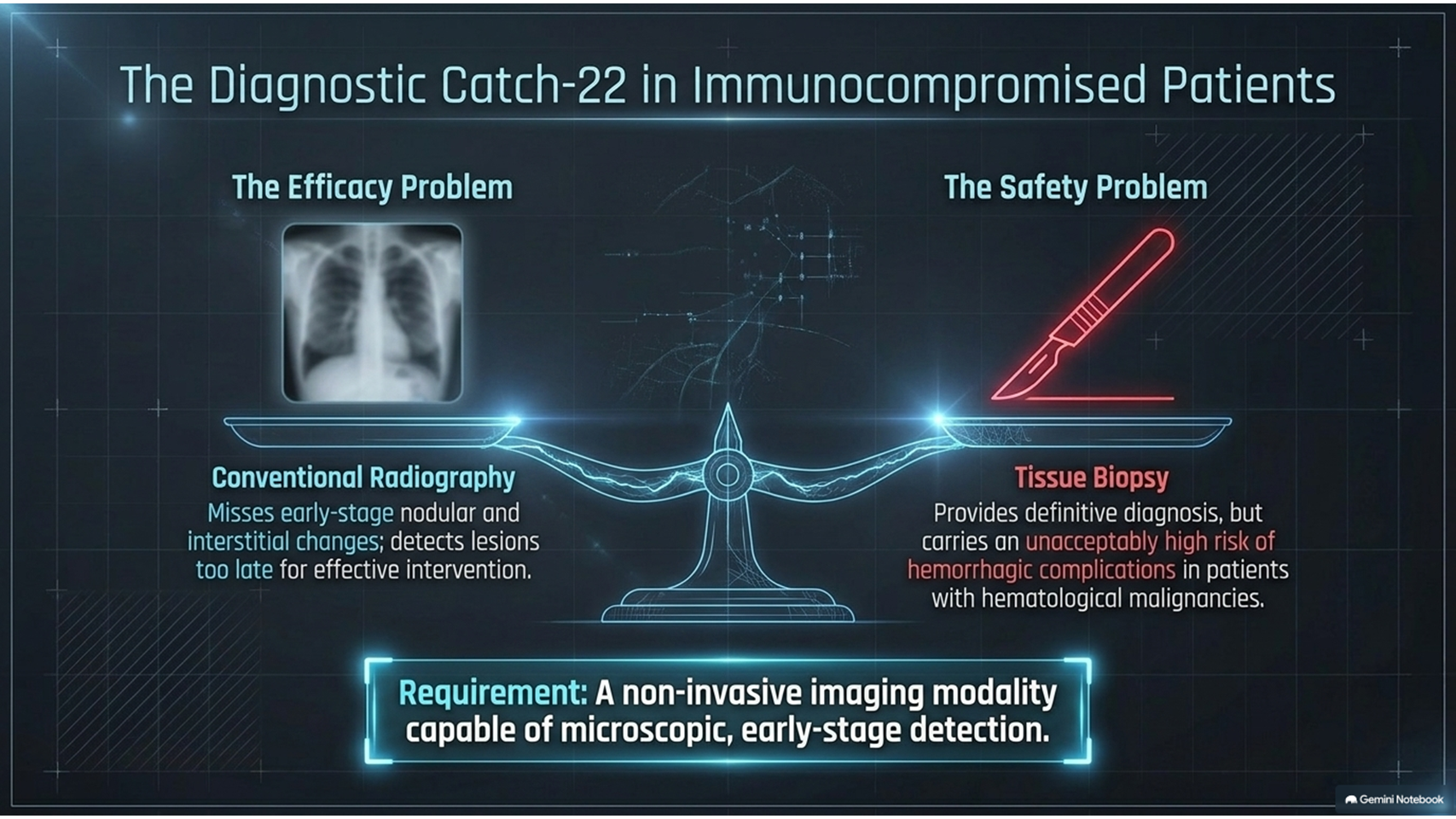
In patients with severe immune suppression, particularly those with granulocytopenia, the clinical signs of invasive pulmonary aspergillosis can become more apparent. In some cases, diagnosis may require biopsy, although biopsy can carry a risk of hemorrhagic complications in patients with hematological malignancies.
Materials and Methods
The study investigated 55 patients with invasive pulmonary aspergillosis. Diagnosis was based on a combination of clinical, immunological, histological, and mycological investigations.
Clinical evaluation included physical examination, clinical and biochemical blood tests, fiber-optic bronchoscopy, and assessment of external respiratory function. Microbiological investigations were performed according to standard procedures.
Additional laboratory investigations included skin testing, measurement of total and specific IgE, examination of sputum and bronchial fluid, and histological analysis using several staining techniques. Aspergillus-specific investigations included detection of antibodies and Aspergillus fumigatus antigen.
The diagnostic imaging protocol included chest radiography and computed non-linear study, or NLS-graphy, of the thorax. The objective of NLS-graphy was to identify and localize abnormal pulmonary parenchyma, detect signs characteristic of fungal involvement, and evaluate relationships between NLS findings, clinical manifestations, and laboratory results.
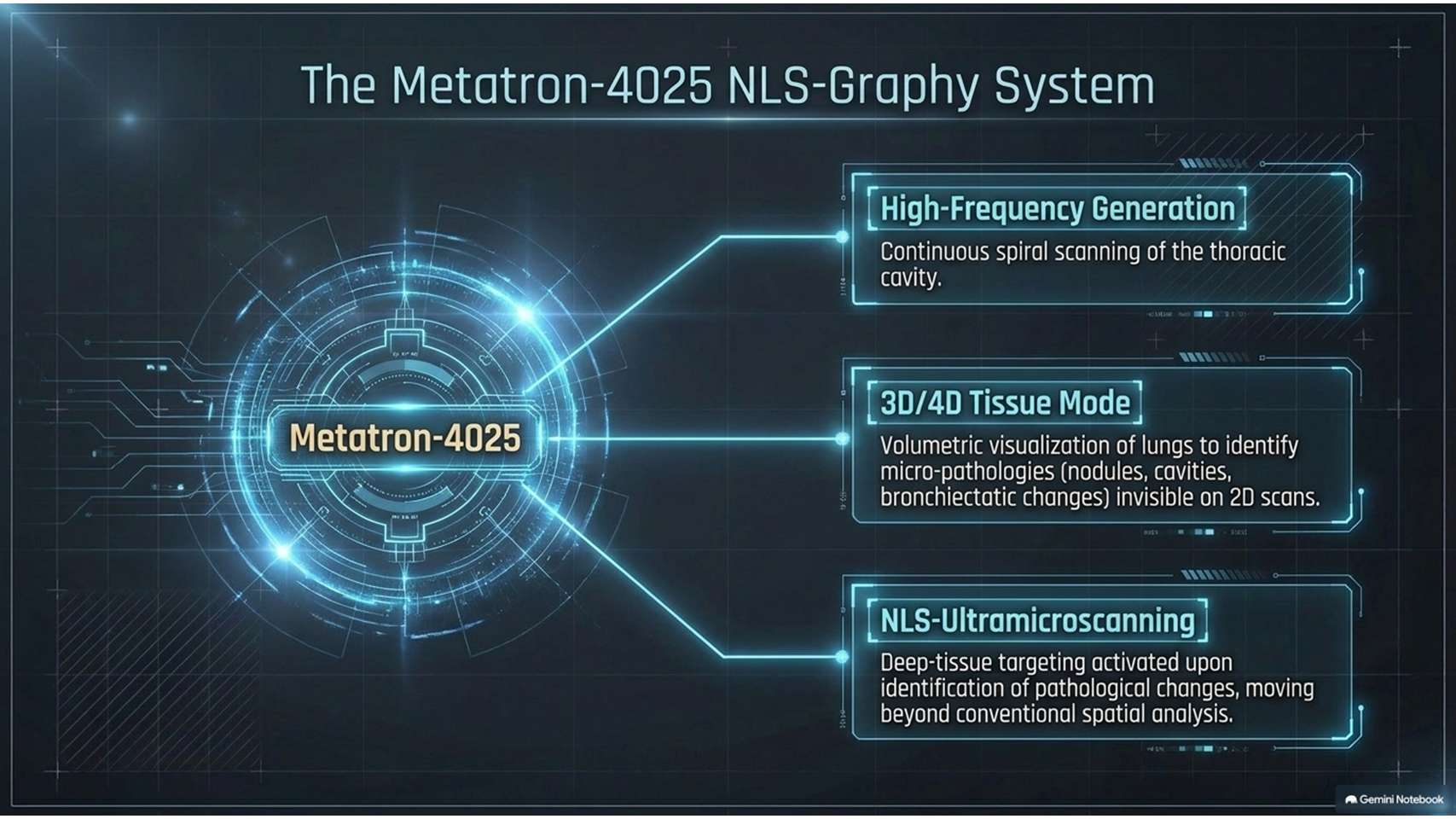
The NLS examination was performed using a Metatron-4025 system equipped with a high-frequency generator, continuous spiral scanning, and software capable of three-dimensional visualization of the lungs. NLS-ultramicroscanning with spectral-entropic analysis was also used to evaluate the spectral similarity of affected lung tissue to an Aspergillus fumigatus reference pattern.
Three-Dimensional NLS Imaging
Three-dimensional NLS imaging was used during the initial thoracic examination. Compared with two-dimensional scanning, the technique was described as allowing better visualization of small pathological lesions, including nodules, cavities, and bronchiectatic changes.
The examination could be extended to 4D Tissue mode with NLS-ultramicroscanning when pathological changes were identified. Repeated NLS examinations were performed in some patients to evaluate disease progression, complications, and responses to therapeutic interventions.
When dissemination of the fungal process was suspected, NLS-ultramicroscanning and spectral-entropic analysis were also performed in other anatomical regions, including the abdominal cavity, brain, and paranasal sinuses.
Clinical Findings in Invasive Pulmonary Aspergillosis
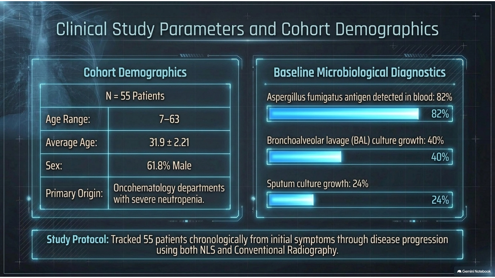
Among the 55 patients with invasive pulmonary aspergillosis, 54 developed the disease in the setting of significantly decreased immunity and severe neutropenia. Most patients were from an oncohematology department.
The patients ranged in age from 7 to 63 years, with an average age of 31.9 ± 2.21 years. Men represented 61.8% of the study population.
Aspergillus fumigatus antigen was detected in blood samples from all patients, with positive results reported in 82% of cases. Cultures of sputum and bronchoalveolar lavage produced Aspergillus fumigatus growth in 24% and 40% of cases, respectively.
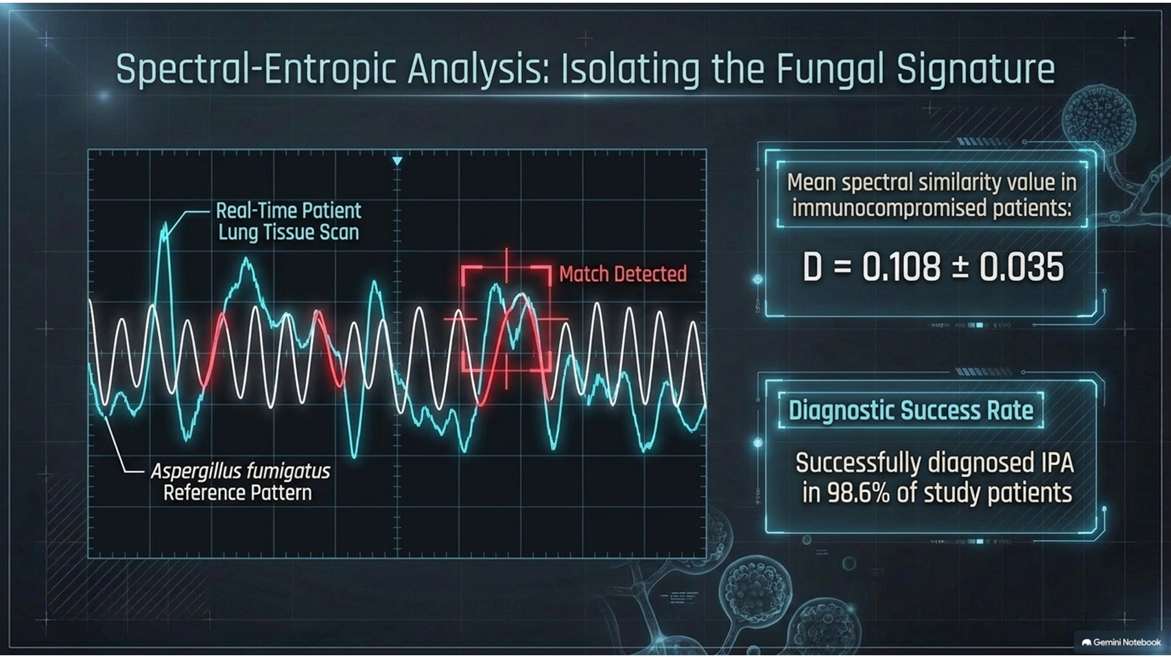
Spectral-entropic analysis diagnosed invasive pulmonary aspergillosis in 98.6% of patients in the study. A high spectral similarity with the Aspergillus fumigatus reference pattern was found in affected lesions.
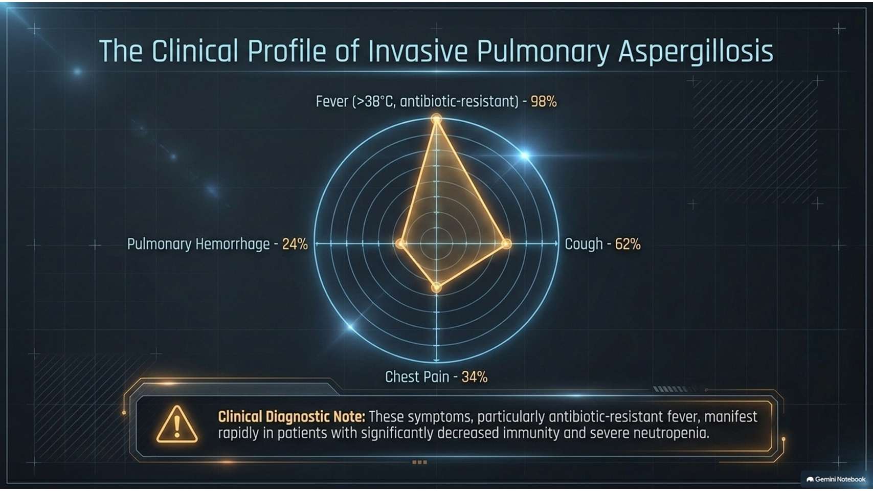
At the early stage of invasive pulmonary aspergillosis, fever above 38°C that was resistant to antibiotic therapy was reported in 98% of cases. Cough occurred in 62%, pulmonary hemorrhage in 24%, and chest pain in 34% of patients.
Dissemination was detected in 5% of patients, and all patients with dissemination had cerebral involvement. Overall, 26% of the patients in the study died.
NLS-Graphy Findings
The NLS findings of invasive pulmonary aspergillosis varied according to the stage of the disease. One of the most frequently observed pulmonary changes was nodular and focal hyperchromogenicity of the pulmonary tissue.
NLS-graphy detected nodular and interstitial changes during the early stages of invasive pulmonary aspergillosis. The study reported that these changes were detected more frequently by NLS than by conventional radiography.
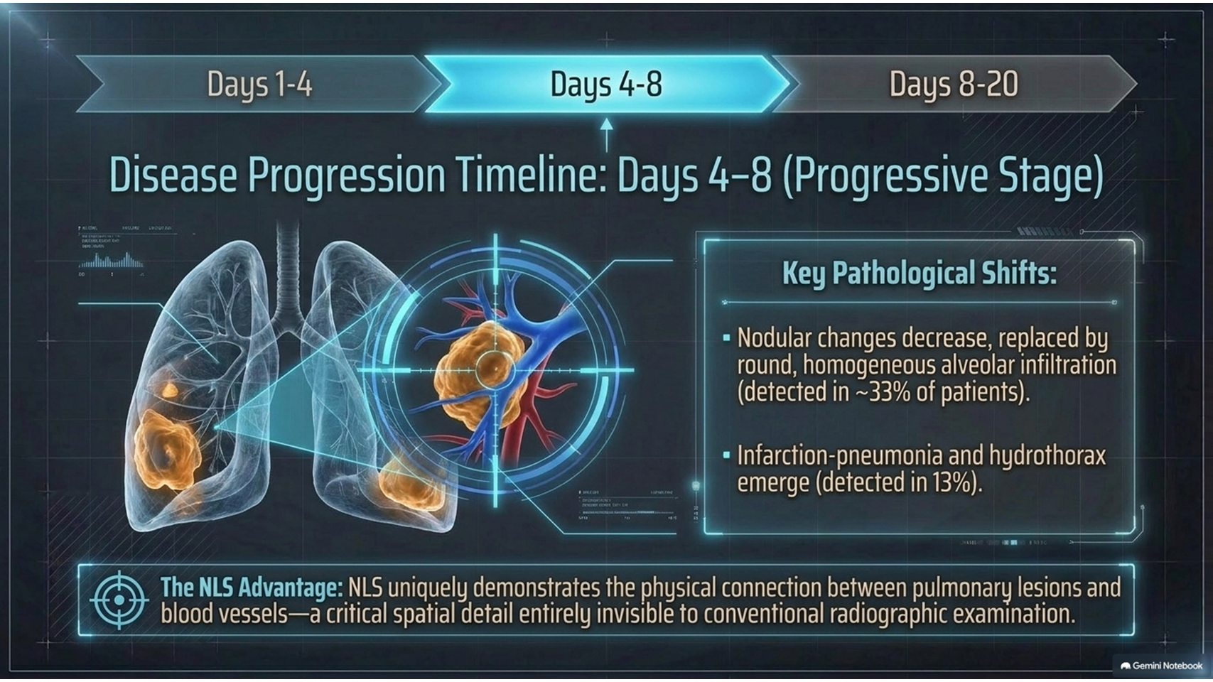
Alveolar infiltration was identified in approximately one-third of patients. Infarction-pneumonia was detected by NLS-graphy in 13% of patients. Hydrothorax was also observed with a similar frequency.
Most nodules, foci, and areas of pulmonary tissue infiltration had a subpleural location. The study also reported that NLS frequently demonstrated a connection between pulmonary lesions and vessels, a finding that was not identified by conventional radiographic examination.
NLS Findings During Different Stages of Disease
The study evaluated three periods following the first appearance of clinical symptoms:
- Days 1–4: Early-stage changes
- Days 4–8: Progressive pulmonary changes
- Days 8–20: Later-stage findings
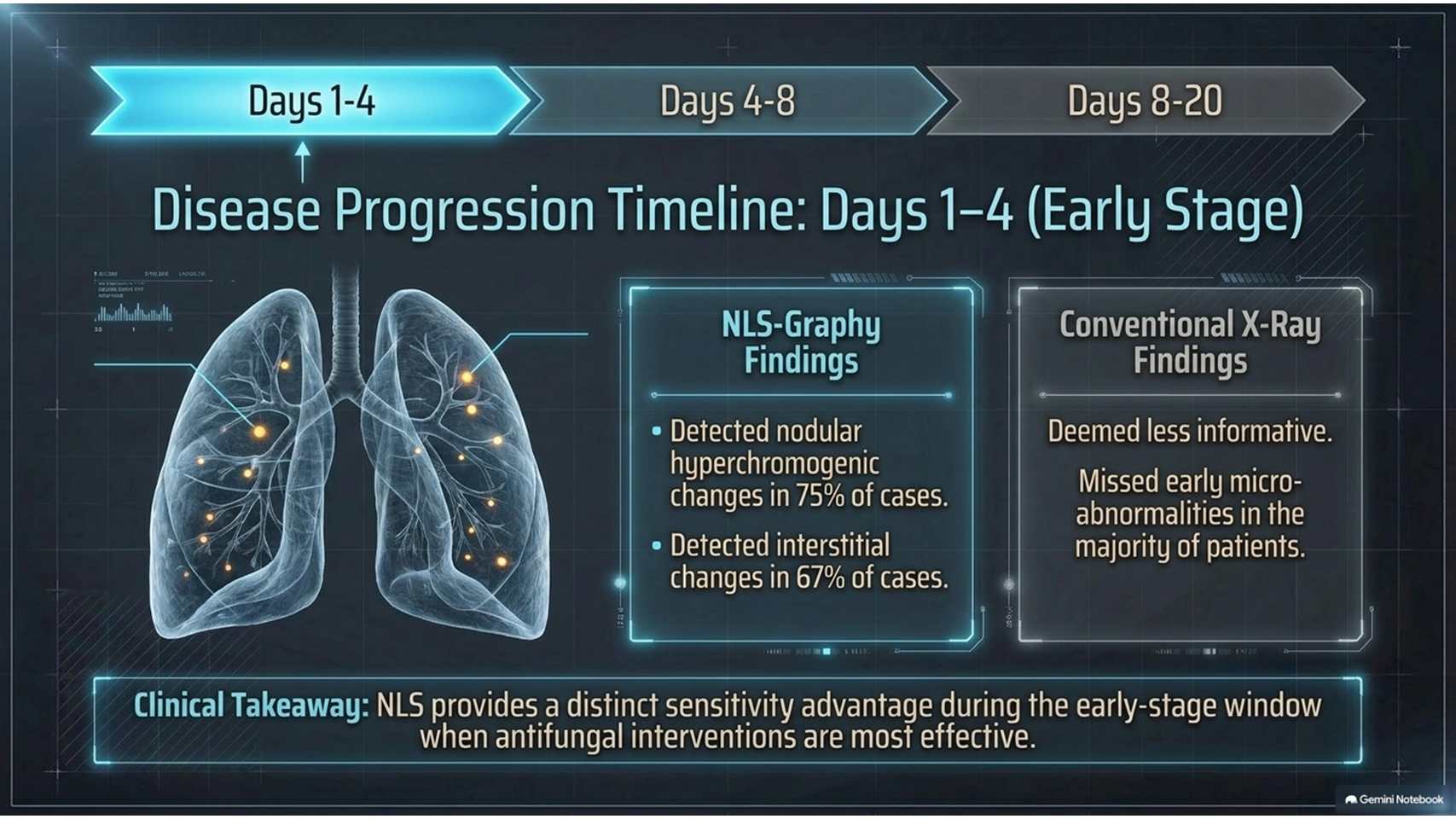
During the first period, the earliest NLS signs of invasive pulmonary aspergillosis were nodular and interstitial hyperchromogenic changes. These were reported in 75% and 67% of cases, respectively.
Conventional radiography was less informative during the early stage, detecting nodular and interstitial changes in fewer patients. NLS therefore demonstrated greater sensitivity for identifying early pulmonary abnormalities in the study.
During the second period, nodular changes became less frequent, while focal areas of alveolar infiltration could be observed. Lesions were often round, relatively homogeneous, subpleural, and connected with vessels.
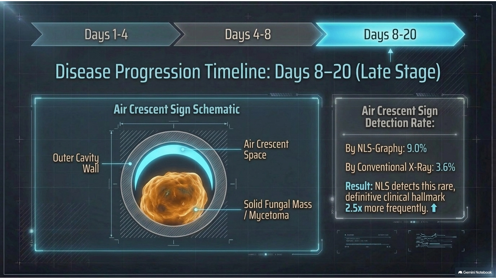
During the third period, from approximately the eighth day onward, the number of detectable changes decreased. However, a characteristic air crescent sign could appear during this stage.
The air crescent sign was described as a cavity containing solid material separated from the cavity wall by a thin crescent-shaped air space. It was detected in 9% of cases by NLS and 3.6% by conventional radiography.
Spectral-Entropic Analysis in Invasive Pulmonary Aspergillosis
Spectral-entropic analysis was an important component of the NLS examination described in the study. In patients with invasive pulmonary aspergillosis, affected lung lesions demonstrated high spectral similarity to the Aspergillus fumigatus reference pattern.
The study reported a high spectral similarity in patients with significant immune suppression. In the investigated group, the mean spectral similarity value was reported as D = 0.108 ± 0.035.
NLS and spectral-entropic analysis were also used to evaluate possible dissemination of invasive pulmonary aspergillosis. In patients with generalized disease, cerebral involvement was detected, and findings were confirmed using NLS and MRI data.
NLS-Graphy Compared With Radiography
According to the study, NLS-graphy was more informative than conventional radiography for detecting pulmonary pathological changes associated with invasive pulmonary aspergillosis.
The reported diagnostic performance of NLS-graphy included:
- Sensitivity: 86%
- Specificity: 75%
- Diagnostic accuracy: 80%
- Statistical significance: p < 0.05
The study therefore described NLS-graphy as a more informative imaging method for the diagnosis of invasive pulmonary mycoses compared with conventional radiography.
Conclusion
Invasive pulmonary aspergillosis is a severe opportunistic fungal infection that primarily affects patients with significant immune suppression. Early recognition is important because the disease can progress rapidly and may disseminate to other organs.
The study found that NLS-graphy could identify pulmonary abnormalities associated with invasive pulmonary aspergillosis more frequently than conventional radiography. Early NLS findings included nodular and interstitial hyperchromogenic changes, while later stages could demonstrate alveolar infiltration, cavitary lesions, and the air crescent sign.
Spectral-entropic analysis also demonstrated high spectral similarity between affected pulmonary tissue and the Aspergillus fumigatus reference pattern.
Based on the reported results, NLS-graphy had a sensitivity of 86%, specificity of 75%, and diagnostic accuracy of 80%. The study concluded that NLS-graphy was more informative than conventional radiography for the diagnosis of invasive pulmonary mycoses.
Important note: The findings and diagnostic performance figures above are those reported in the supplied source. They are presented as study findings rather than as independent clinical recommendations.




No comment