

Introduction
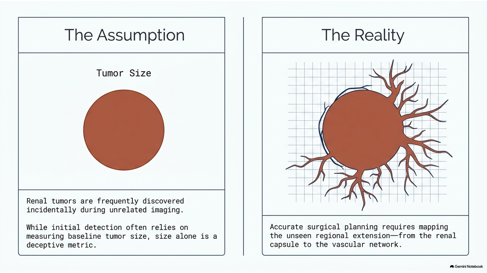
Renal cancer can be challenging to diagnose and stage because it may develop without specific clinical symptoms during its early stages. Many renal tumors are discovered incidentally during imaging examinations performed for unrelated medical conditions. Early detection of a renal tumor is important because accurate assessment of tumor size, local extension, vascular involvement, and regional metastasis can influence treatment planning.
3D NLS-Graphy for renal cancer staging was investigated as an imaging approach for evaluating renal tumors and determining the regional extent of renal cancer. The objective of 3D NLS-Graphy for renal cancer staging was to identify or clarify the stage of the primary tumor and, in selected cases, assist with the differential assessment of detected renal neoplasms.
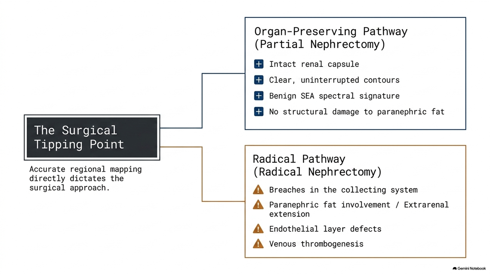
Accurate renal cancer staging is particularly important when minimally invasive and organ-preserving surgical techniques are being considered. Determining whether a renal tumor remains confined to the kidney or extends into the renal capsule, surrounding tissues, collecting system, renal vein, or inferior vena cava can affect the choice of surgical treatment.
Materials and Methods
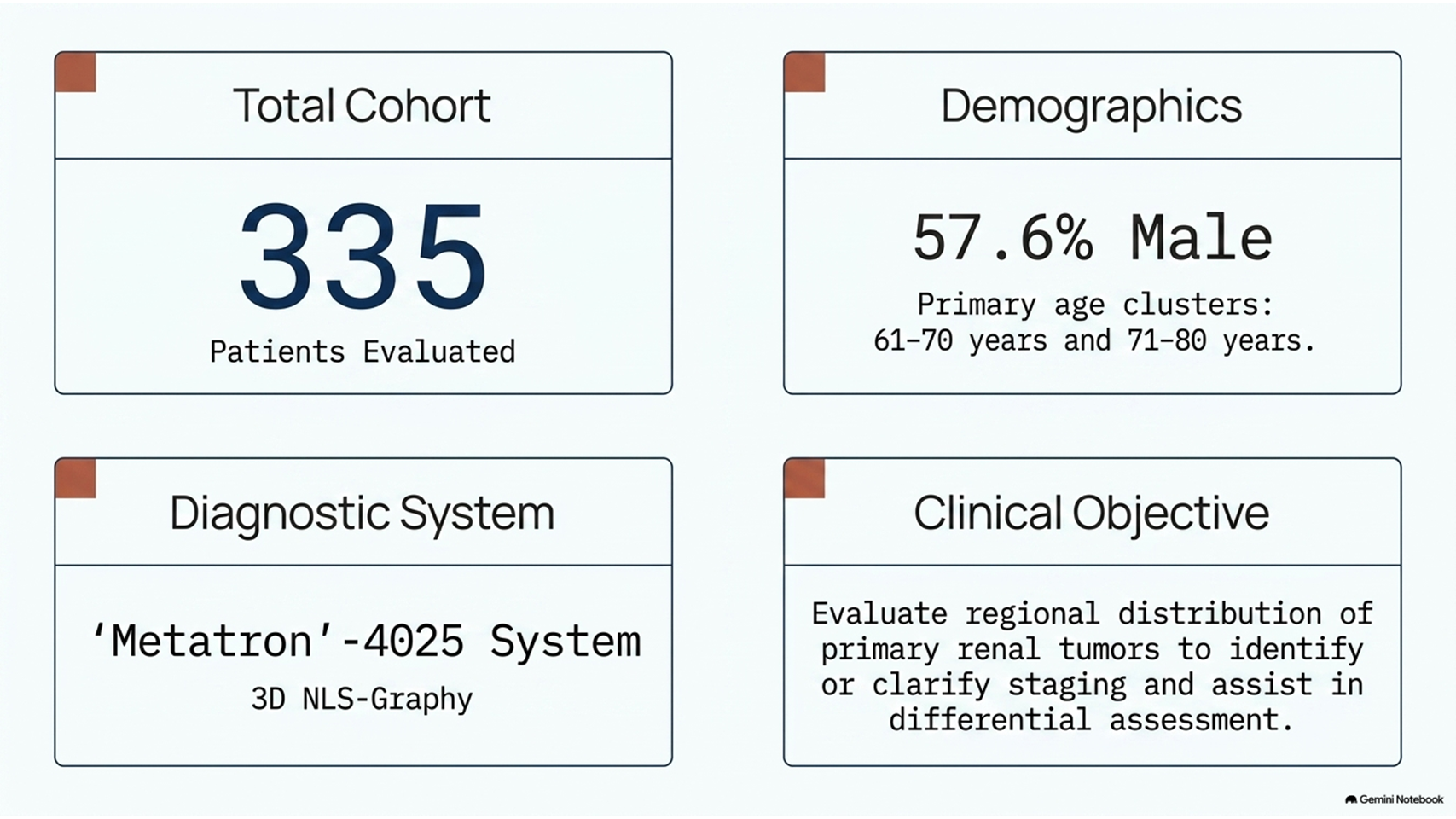
A total of 335 patients with renal cancer were evaluated in the reported study. Men represented 57.6% of the patient population. The largest age groups were patients aged 61–70 years and 71–80 years.
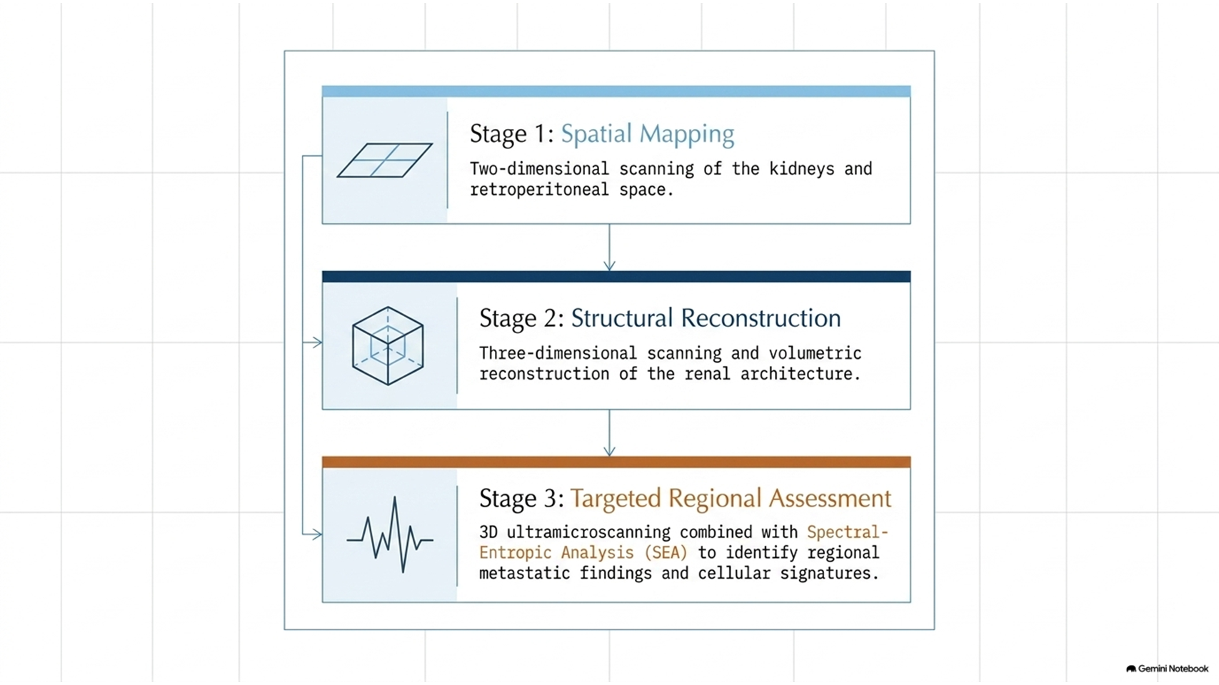
The NLS examination was performed using the “Metatron”-4025 diagnostic system. In 213 patients, pre-surgical NLS examination was performed using a standard protocol that included two consecutive stages:
- Two-dimensional scanning of the kidneys and retroperitoneal space.
- Three-dimensional scanning and reconstruction of the kidneys and retroperitoneal space.
In addition, a proportion of patients underwent 3D ultramicroscanning with spectral-entropic analysis (SEA).
The purpose of 3D NLS-Graphy for renal cancer staging was to evaluate the regional distribution of the primary renal tumor. The assessment focused on tumor size, local invasion, vascular involvement, and regional metastatic findings.
Results of 3D NLS-Graphy for Renal Cancer Staging
The reported NLS findings were organized according to several characteristics of renal cancer progression, including the direction of primary tumor invasion, vascular involvement, and regional metastasis.
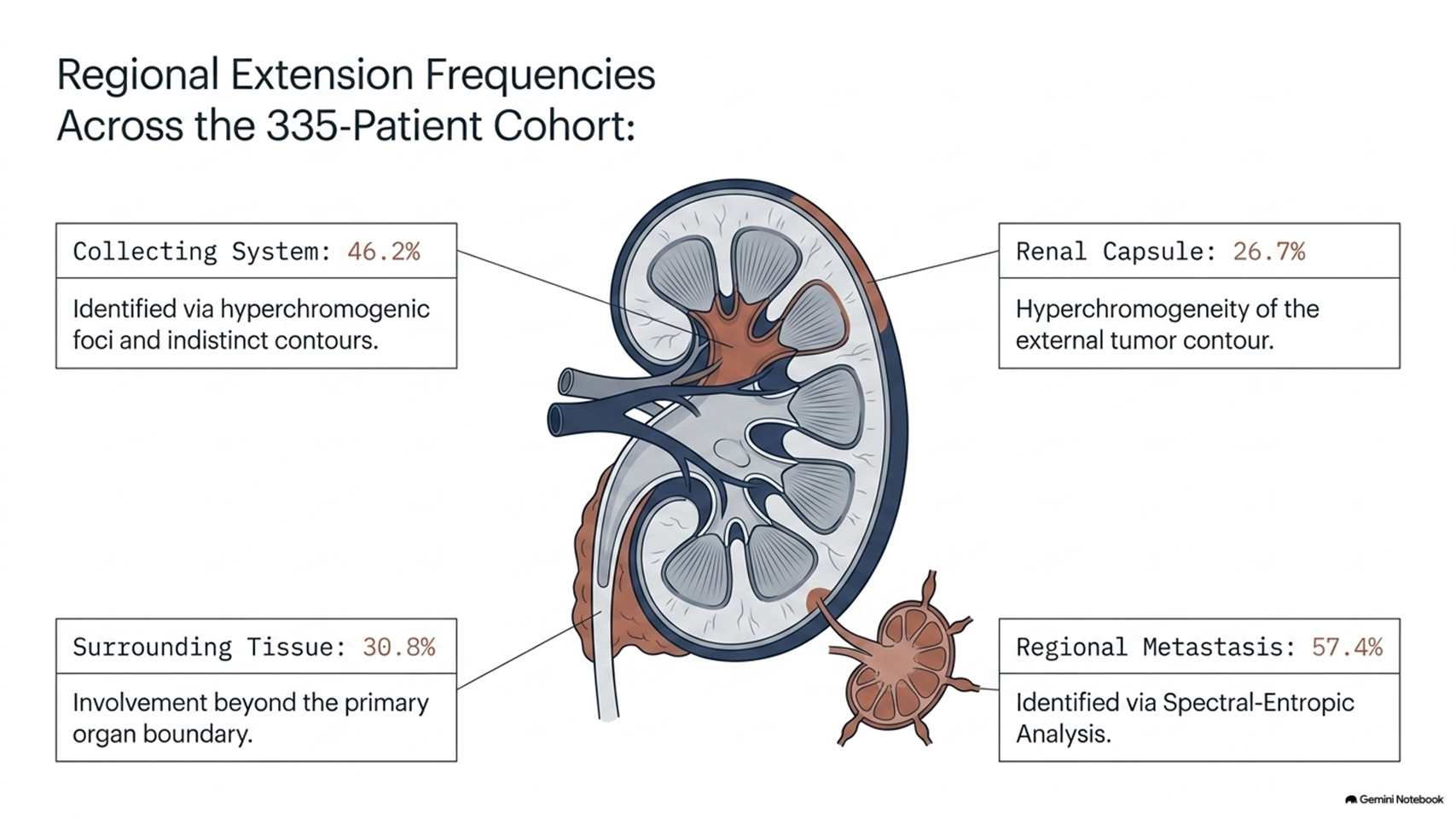
One of the most frequently reported findings was tumor invasion involving the structural elements of the kidney’s collecting system. Hyperchromogenic foci were identified in 46.2% of cases, while indistinct contours of the collecting system were also observed.
Another important group of findings involved extension of the renal tumor beyond the kidney. Findings associated with surrounding tissue involvement were reported in 30.8% of cases. Hyperchromogeneity of the external tumor contour or capsule was reported in 26.7% of patients.
SEA findings associated with regional metastasis were reported in 57.4% of the studied patients.
These findings suggest that 3D NLS-Graphy for renal cancer staging was used in the reported study to evaluate more than the size of the primary renal tumor. The examination was also used to assess possible local tumor extension, surrounding tissue involvement, vascular changes, and metastatic findings.
Renal Tumor Size and Local Tumor Extension
The reported frequency of several local invasion findings was related to the size of the primary renal tumor.
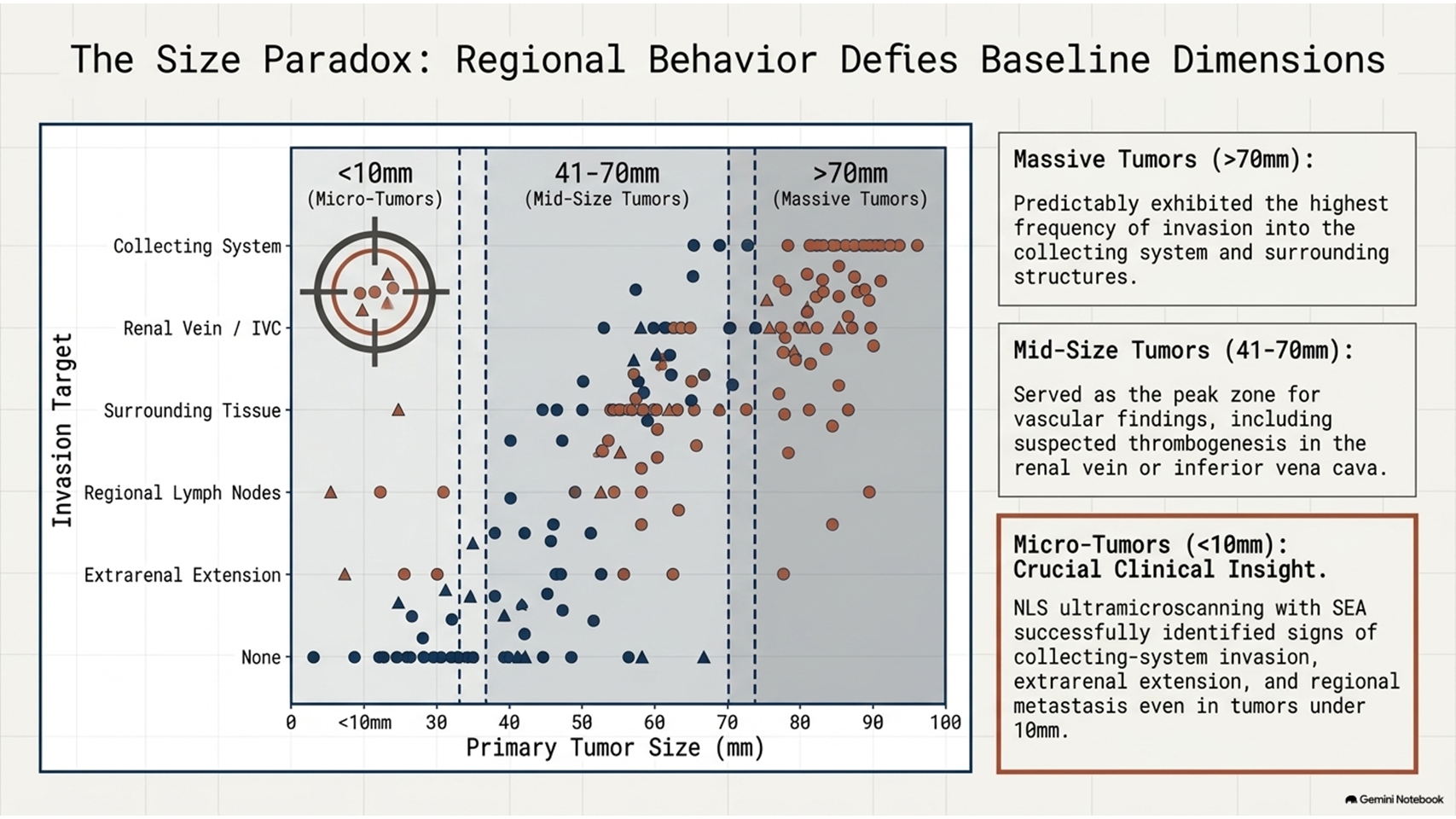
The highest frequency of findings associated with invasion of the collecting system and surrounding structures was observed in tumors larger than 70 mm.
Vascular findings, including suspected thrombogenesis or invasion involving the renal vein or inferior vena cava, were reported more frequently in tumors measuring between 41 and 70 mm. Findings associated with other vascular changes were also reported in smaller renal tumors.
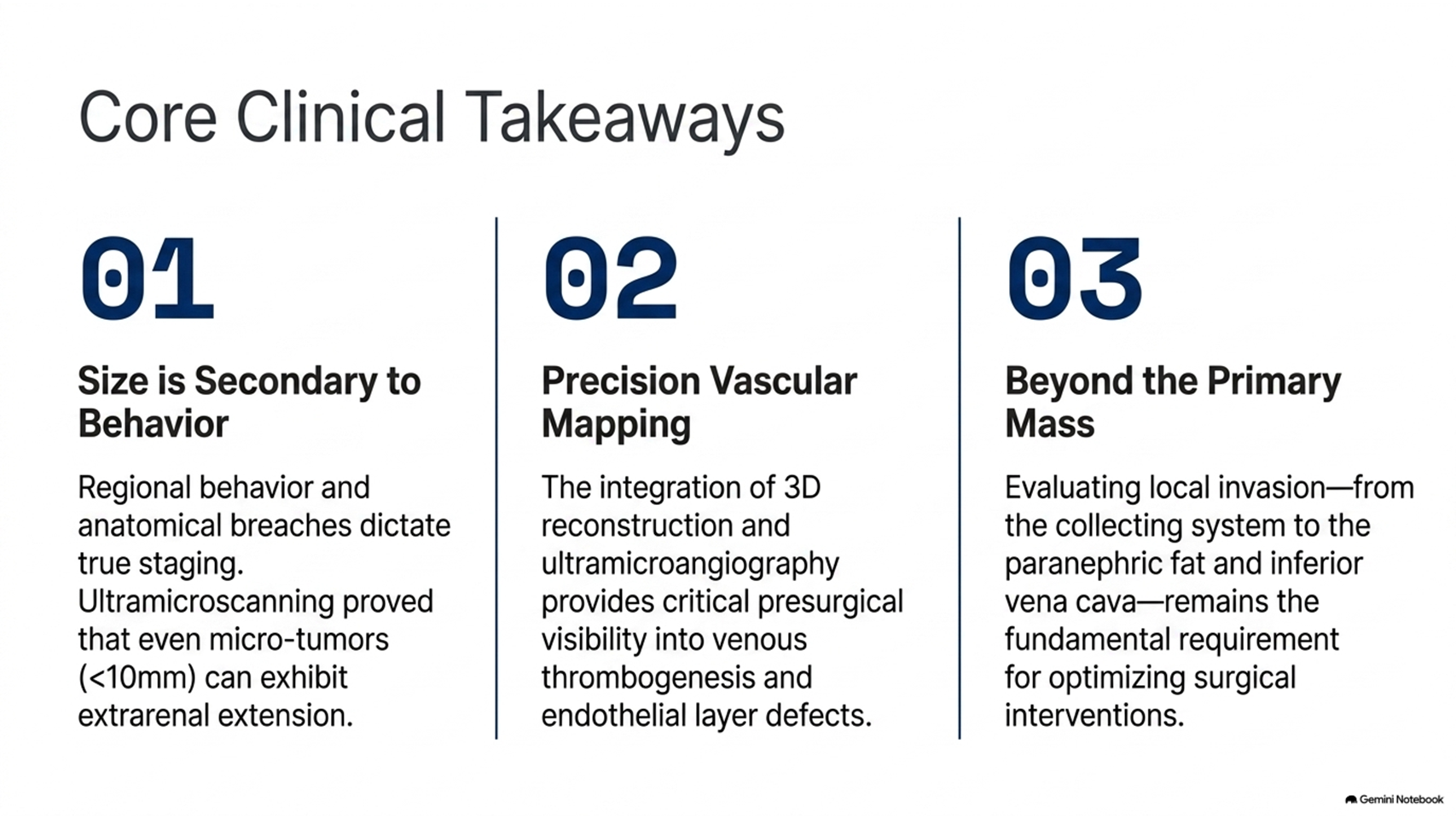
An important observation in the study was the reported ability of NLS ultramicroscanning with SEA to identify signs of regional tumor extension in some tumors smaller than 10 mm. In these individual cases, findings included collecting-system invasion, extrarenal extension, and regional metastatic findings.
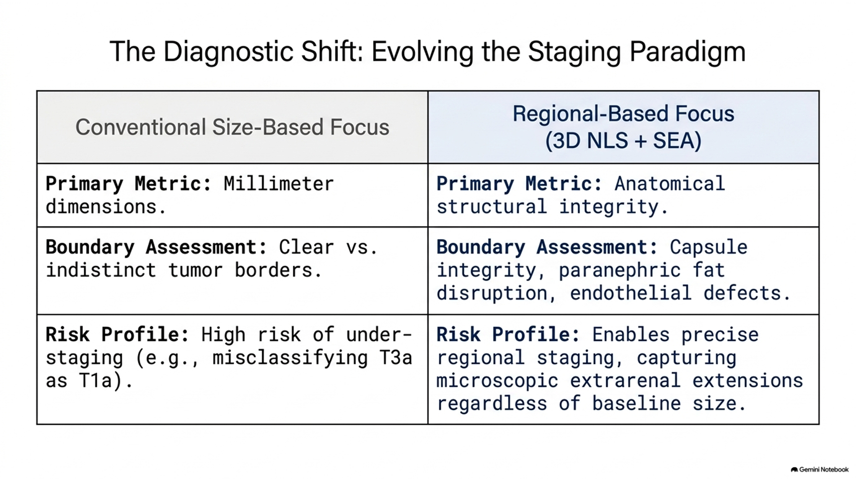
Therefore, the reported application of 3D NLS-Graphy for renal cancer staging extended beyond conventional tumor-size assessment and focused on evaluating the regional behavior of renal tumors.
Clinical Observation 1: Renal Cancer and Collecting System Invasion
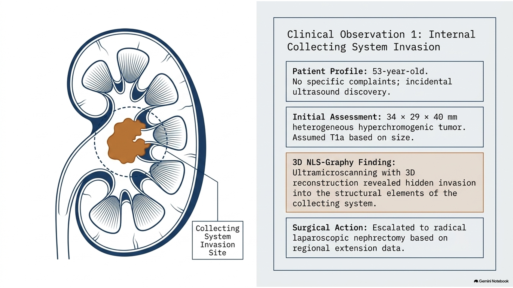
A 53-year-old patient was referred for urological evaluation without specific complaints. A prophylactic ultrasound examination identified a tumor in the right kidney.
Two-dimensional NLS examination identified a heterogeneous hyperchromogenic renal parenchymal tumor measuring approximately 34 × 29 × 40 mm.
Based on tumor size, the lesion was initially classified as T1a. To further evaluate the regional extension of the renal tumor, NLS ultramicroscanning with SEA and three-dimensional reconstruction were performed.
The 3D examination demonstrated findings interpreted as invasion of the primary renal tumor into structural elements of the collecting system.
Based on the reported imaging findings, radical laparoscopic nephrectomy was selected.
This clinical observation illustrates how 3D NLS-Graphy for renal cancer staging was used to provide additional information about tumor extension that was not apparent from tumor size alone.
Clinical Observation 2: Renal Tumor Extension Into Surrounding Tissue
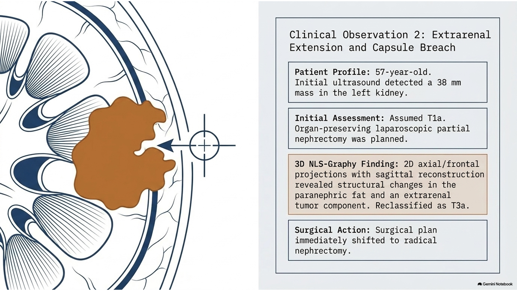
A 57-year-old patient was evaluated after an ultrasound examination detected a mass in the left kidney. A subsequent ultrasound examination identified a renal parenchymal tumor measuring approximately 38 mm.
The lesion was initially considered a T1a renal tumor, and an organ-preserving laparoscopic partial nephrectomy was considered.
Additional NLS examination was performed to clarify the tumor stage and determine the most appropriate surgical approach.
Two-dimensional NLS images in axial and frontal projections, together with sagittal reconstruction, demonstrated findings interpreted as extrarenal extension. These findings included structural changes in the paranephric fat and an extrarenal tumor component.
Following NLS ultramicroscanning with SEA, the tumor was classified in the reported study as T3a.
The surgical plan was subsequently changed to radical nephrectomy.
This case demonstrates the potential role attributed to 3D NLS-Graphy for renal cancer staging in differentiating a tumor that appears limited in size from a tumor with suspected extension beyond the kidney.
Renal Cancer and Vascular Invasion
Vascular involvement is an important consideration in renal cancer staging because renal tumors may extend into the renal vein or inferior vena cava.
The reported study described NLS ultramicroangiography and three-dimensional reconstruction as methods used to evaluate possible thrombogenesis and vascular involvement.
Reported findings included intraluminal thrombotic masses and defects involving the endothelial layer.
The assessment of renal vein and inferior vena cava involvement can be particularly important before renal cancer surgery because vascular extension may influence surgical planning.
For this reason, evaluation of vascular structures represented an important component of 3D NLS-Graphy for renal cancer staging in the reported cases.
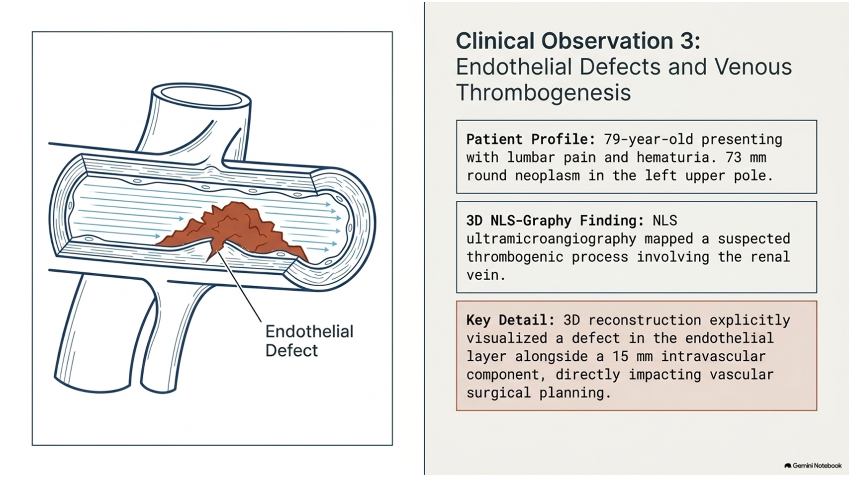
Clinical Observation 3: Renal Vein Involvement
A 79-year-old patient presented with lumbar pain and episodes of hematuria. Ultrasound examination identified a tumor involving the upper pole of the left kidney.
NLS examination demonstrated a round renal neoplasm measuring approximately 73 mm.
NLS ultramicroscanning identified a suspected thrombogenic process involving the renal vein. A defect in the endothelial layer was described, with an intravascular component measuring approximately 15 mm.
The vascular finding was clearly visualized following three-dimensional reconstruction.
This case highlights the reported use of 3D NLS-Graphy for renal cancer staging to evaluate possible vascular involvement in a patient with a large renal tumor.
Differential Assessment of Renal Tumors
In addition to renal cancer staging, the reported study investigated the use of spectral-entropic analysis for differential assessment of renal parenchymal masses.
According to the study, SEA was used to differentiate potentially malignant and benign renal lesions. The reported results were compared with biopsy findings, with a reported confirmation rate of 98.2%.
However, these historical findings should be interpreted in the context of the study design and should not be considered equivalent to current validated diagnostic standards without independent clinical evidence.
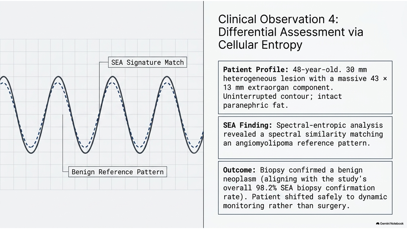
Clinical Observation 4: Suspected Benign Renal Neoplasm
A 48-year-old patient underwent ultrasound examination, which demonstrated a heterogeneous round lesion in the right kidney.
The lesion measured approximately 30 mm, with an extraorgan component measuring up to 43 × 13 mm. The contour was described as clear and uninterrupted, and no structural damage of the paranephric fat was identified.
Because the ultrasound findings required further differential assessment, SEA was performed.
The reported spectral similarity of the lesion to an angiomyolipoma reference pattern supported a benign interpretation. Biopsy subsequently confirmed a benign renal neoplasm, and dynamic monitoring was recommended.
This clinical observation demonstrates another reported application of NLS and SEA in the assessment of renal tumors in addition to 3D NLS-Graphy for renal cancer staging.
Importance of Regional Renal Cancer Staging
Renal cancer staging involves more than measuring the primary tumor. Evaluation of local invasion, surrounding tissues, collecting-system involvement, vascular extension, and regional metastasis can provide important information for treatment planning.
The reported study focused on these characteristics and proposed 3D NLS-Graphy for renal cancer staging as a method for evaluating regional tumor extension.
The study particularly emphasized the identification of suspected invasion into:
- The renal collecting system
- The renal capsule
- Paranephric fat
- Surrounding tissues
- The renal vein
- The inferior vena cava
- Regional lymphatic structures
Assessment of these structures may be relevant when determining whether an organ-preserving approach or radical surgery is appropriate.
3D NLS-Graphy for Renal Cancer Diagnosis and Staging
The reported findings indicate that 3D NLS-Graphy for renal cancer staging was primarily investigated as a pre-surgical assessment technique.
The combination of two-dimensional imaging, three-dimensional reconstruction, ultramicroscanning, and spectral-entropic analysis was reported to provide additional information about renal tumor morphology and regional extension.
In particular, the study emphasized the potential ability of NLS examination to identify suspected tumor extension even when conventional ultrasound primarily demonstrated a relatively small renal mass.
Nevertheless, modern renal cancer diagnosis and staging generally rely on validated imaging techniques and pathological assessment when clinically indicated. Therefore, the historical NLS findings presented in this article should be considered within the context of the original study rather than as a replacement for established contemporary diagnostic methods.
Conclusion
The reported study evaluated 3D NLS-Graphy for renal cancer staging in 335 patients with renal cancer.
According to the study, NLS examination was used to evaluate the size and regional extension of renal tumors, including suspected involvement of the collecting system, renal capsule, paranephric fat, surrounding tissues, renal vein, inferior vena cava, and regional metastatic structures.
The study reported that 3D NLS-Graphy for renal cancer staging could provide additional information during pre-surgical assessment, particularly when evaluating local tumor extension and suspected vascular involvement.
The study also reported the use of spectral-entropic analysis for differential assessment of renal masses, with biopsy confirmation reported in 98.2% of cases.
Overall, the historical findings presented in this study describe 3D NLS-Graphy for renal cancer staging as an investigated imaging approach for evaluating renal tumor characteristics and regional disease spread. Further assessment against modern imaging and diagnostic standards is necessary to determine its current clinical relevance.
References
- Novick AC, Campbell SC. Renal tumors. In: Walsh PC, Retik AB, Vaughan ED. Campbell’s Urology. Philadelphia, 2002.
- Pantuck AJ, Zisman A, Belldegrun AS. The changing natural history of renal cell carcinoma. Journal of Urology. 2001.
- Bullinger S, Bonnet T. Achievements of ray diagnostics – a progress in urology. Materials of the 2nd International Congress on Ray Diagnostics and Therapy. Prague, 2009.
- Legard S. Certain methods of renal cancer remote treatment results improving. Oncourology. 2009.
- Kvon D. Comparative analysis of results achieved with radical nephrectomy carried out by open and laparoscopic approaches. Oncourology. 2009.
- Nesterova VI, Shaposhnikov LV, Yankina LA, Kozhemyakin OR. Application of NLS-diagnostics in oncology: New trends and prospects. NLS-Diagnostics in Medicine: Prospects of Development. Moscow, 2010.




No comment